Excess iron in vital organs, even in mild cases of iron overload, increases the risk for liver disease (cirrhosis, cancer), heart attack or heart failure, diabetes mellitus, osteoarthritis, osteoporosis, metabolic syndrome, hypothyroidism, hypogonadism, numerous symptoms and in some cases premature death.
Researchers in China recently evaluated the changes of ferritin levels in the brain’s cerebrospinal fluid of patients with amyotrophic lateral sclerosis (ALS). They found that ALS patients exhibited significantly increased levels of ferritin, an iron-based protein, a result that indicates ferritin has value as a biomarker and could be used for diagnostic and monitoring purposes in ALS.
The study, ‘’Elevated levels of ferritin in the cerebrospinal fluid of amyotrophic lateral sclerosis patients,’’ was published in Acta Neurologica Scandinavica.
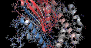
Many studies have reported that ALS is caused mainly by genetic mutations, which induce the progressive deterioration and loss of motor neurons. Some recent findings, however, have suggested that metal toxicity and changes in ferritin, a blood cell protein that binds to iron and stores it inside the cells, could be risk factors for the development of ALS. That is because iron overload has been associated with DNA/RNA damage.
A few other published investigations pointed to increased levels of iron and ferritin in the serum of patients with ALS. However, little is known about the levels of iron and ferritin in the brain’s cerebrospinal fluid (CSF) of ALS patients.
In this study, the researchers evaluated changes in levels of several iron proteins in CSF, including ferritin heavy chain (FHC), ferritin light chain (FLC), and transferrin. FHC and FLC are subunits (parts) of the ferritin protein, while transferrin is a blood plasma protein that controls the level of free iron; high transferrin levels usually are associated with iron deficiency.
For comparative purposes, these species also were measured in serum and compared to those obtained in the brain.
Serum and CSF samples were collected from 54 ALS patients and 46 controls subjects. Levels of iron-based proteins then were estimated in both serum and CSF for both ALS patients and controls and the data were analyzed and compared.
The results suggested ALS patients have significantly higher levels of FHC and FLC in CSF when compared to controls. Furthermore, these levels were found to rise as the disease progresses, but reduce with disease duration. For transferrin, high levels were detected in the serum of ALS patients, but no specific correlation to disease progression was noticed. Finally, as the disease increased in severity, the FHC and FLC in CSF rapidly rose.
“Our study determined that FHC and FLC could be identified in CSF and that their measurements may in fact be useful in disease differentiation and evaluating progression in patients with ALS,’’ the authors concluded.
“As the levels of these proteins in CSF differed from those in the serum, CSF measurements may be more reliable than serum measurements with respect to diagnosing the disease and evaluating disease progression. This study was unable to determine whether changes in the levels of FHC and FLC in CSF were indicators of disease occurrence or outcome,” they added.
NOVEMBER 11, 2016 BY MALIKA AMMAM, PHD IN NEWS.
https://alsnewstoday.com/2016/11/11/study-finds-als-patients-with-elevated-levels-of-iron-based-proteins-in-brain
